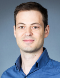
Prof. Michael Doube is Clinical Associate Professor in Anatomy at the Department of Infectious Diseases and Public Health.
He earned a veterinary degree at Massey University, New Zealand and a PhD at Queen Mary, University of London, on early changes in the equine third metacarpal bone in the same site as later condylar fracture. During a postdoc at Imperial College London's Department of Bioengineering, in collaboration with the RVC's Structure and Motion Laboratory, Michael investigated scaling of bone microstructure and gross dimensions in relation to animal size. To accomplish this research, Michael started the BoneJ software project, which brought together existing and new programs for bone image analysis. After a stint at the Light Microscopy Facility of the Max Planck Institute of Molecular Cell Biology and Genetics in Dresden, Michael returned to London to take up a lectureship within The Royal Veterinary College's Department of Comparative Biomedical Sciences.
Prof. Doube's research concentrates on imaging and bioimage informatics of skeletal tissues.
Michael continues to work on BoneJ, now in its second major iteration (BoneJ2), supported by funding from the Wellcome Trust, the Royal Society, and BBSRC. He is investigating comparative skeletal physiology and anatomy, using imaging at the organ, tissue and cellular levels. Michael is an active member of the ImageJ and Fiji community, and supports BoneJ users via the ImageJ forum. Michael is a member of the Bone Research Society, a Fellow of the Royal Microscopical Society and an Associate Editor for Royal Society Open Science.
For an up-to-date list of publications and citations, please see CityU Scholars.
Secondary osteons scale allometrically in mammalian humerus and femur.
Felder AA, Phillips C, Cornish H, Cooke M, Hutchinson JR, Doube M.
R Soc Open Sci. 2017 Nov 8;4(11):170431. doi: 10.1098/rsos.170431
Structure Model Index Does Not Measure Rods and Plates in Trabecular Bone.
Salmon PL, Ohlsson C, Shefelbine SJ, Doube M.
Front Endocrinol (Lausanne). 2015 Oct 13;6:162. doi: 10.3389/fendo.2015.00162
Trabecular bone scales allometrically in mammals and birds.
Doube M, Klosowski MM, Wiktorowicz-Conroy AM, Hutchinson JR, Shefelbine SJ.
OProc Biol Sci. 2011 Oct 22;278(1721):3067-73. doi: 10.1098/rspb.2011.0069
BoneJ: Free and extensible bone image analysis in ImageJ.
Doube M, Kłosowski MM, Arganda-Carreras I, Cordelières FP, Dougherty RP, Jackson JS, Schmid B, Hutchinson JR, Shefelbine SJ.
Bone. 2010 Dec;47(6):1076-9. doi: 10.1016/j.bone.2010.08.023
Variations in articular calcified cartilage by site and exercise in the 18-month-old equine distal metacarpal condyle.
Doube M, Firth EC, Boyde A.
Osteoarthritis Cartilage. 2007 Nov;15(11):1283-92 doi:10.1016/j.joca.2007.04.003
