![]()
|
|
|
|
|
|
Design and applications of psuedo three-dimensional cell-culture substrates |
|||||
|
Representative SEM image of cells grown in a pore. |
In vitro experiments rely heavily on tissue culture, e.g., to study the cell differentiation, proliferation and function, etc. These in vitro experiments usually involve flat culture substrates, e.g., through the use of Petri-dishes and flasks, and are thus convenient for routine growth of cells. However, such conventional cell culture will normally generate only two-dimensional (2D) cell monolayers. Such 2D monolayers will lead to highly abnormal geometric and mechanical pressures on many types of cells, which are far from the realistic conditions and complexities of three-dimensional (3D) tissues. We explored the fabrication of three-dimensional (3D) substrates by creating micrometer-size pores on polyallyldiglycol carbonate (or PADC) polymer films through irradiation of the film by alpha particles and subsequent chemical etching. HeLa cells cultured on these 3D substrates were observed using scanning electron microscope. Multiple directions and multiple layers of HeLa cells were observed to have grown in the pores, with normal nuclei and cell membranes as well as good cell spreading. For the cells cultured in 3D substrates with or without additional small pores, no significant differences were observed between their vinculin expression profiles, which was in contrast to the observation made for cells cultured on 2D substrates showing that small pores could enhance vinculin expression. The presence of the large pores and/or the enhanced biocompatibility of the substrate in the present experiments might be the reasons. The protrusions of cells were confined by the small pores, which was similar to the observation made for cells cultured on 2D substrates. |
||||
|
|||||
|
Effects of substrate topography on cell behavior |
|||||

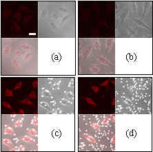
Superposition of HeLa cells and track-etch pits generated from irradiation of 3 MeV alpha particles for 3 h, etched for 3 h in 6.25 N aqueous NaOH at 70 oC and then for 5 min in 1 N NaOH/ethanol at 40 oC. |
It is well established that pores introduce topographies onto the substrates, while substrate topographies will control the nature and degree of cell-cell and cell-matrix interactions and determine the morphology and functional induction of cultured cells in vivo. However, it would be sometimes difficult to separate the relative contribution of topography and porosity from pores. We explored the feasibility of using pits created on the surface of a polymer
(polyallyldiglycol carbonate or
PADC) by alpha-particle irradiation and
subsequent chemical etching to study the substrate topographical effects on
behaviors of cells (HeLa cervix cancer cells). The pits were purposely not etched-through, so that
topographical effects
alone (excluding porosity
effects) can be studied. |
||||
|
|||||

|
Nuclear Radiation Unit |
Page last modified on 13-Jun-2012 Privacy Policy - Copyright - Disclaimer
|